
2D Echo
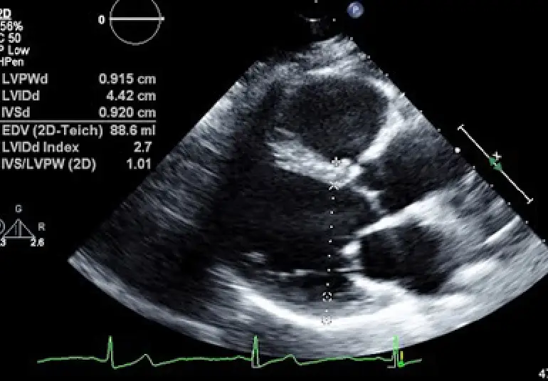
When diagnosing heart conditions in a safe and effective manner, 2D Echo (2D Echocardiography) is one of the most valuable resources of any modern cardiology practice. It relies on high-frequency sound waves (ultrasound) to create real-time pictures of the heart so that doctors can assess its shape and function, all this without taking any invasive procedure into action.
A major adaptation of this is the Fetal 2D Echo, a special exam to evaluate the heart of a developing baby in utero. Early identification of congenital heart illness within the womb can be life-saving.
Let’s dive into what these exams are, how they’re conducted, and why they’re so important.
What is a 2D Echo?
2D Echocardiography is an innocent, non-invasive test that uses ultrasound to produce moving pictures of the heart. It helps doctors visualize the heart’s chambers, valves, walls, and blood circulation.
Is it necessary for whom?
- Those who are suffering from symptoms like chest pain, breathlessness, or palpitations
- Those with a history of heart disease or abnormal ECG
- Those who have hypertension or diabetes
- Those who are being evaluated for pre-operative tests
What Does a 2D Echo Diagnose?
- Disorders of the valves (e.g., mitral regurgitation, aortic stenosis)
- Congenital heart disease
- Damage to the heart muscle (cardiomyopathy)
- Fluid around the heart (pericardial effusion)
- Heart function after myocardial infarction
- Pulmonary hypertension
The Procedure:
The test takes only 20–30 minutes. Gel is put on the chest and a probe, or transducer, is rolled around to capture images. It’s completely safe and doesn’t involve any radiation.
What is Fetal 2D Echo?
Fetal 2D Echocardiography is a specific pregnancy ultrasound, typically performed at 18–24 weeks of pregnancy, to examine the unborn child’s heart in more detail.

Why Fetal 2D Echo is performed?
- Family history of congenital heart disease
- Abnormal result in a routine fetal scan
- Maternal conditions like diabetes or infection
- Increased nuchal translucency or abnormal screening test
Benefits of Fetal 2D Echo
- Early Detection: Identifies structural heart defects like septal defects, valve defects, and complex congenital malformations.
- Better Planning: Allows doctors and parents to plan the birth and treatment of a heart-defective baby.
- Greater Survival: Allows for early intervention or surgery soon after birth, which can provide a dramatic improvement in survival and health of the infant.
Procedure
Performed essentially like a routine pregnancy ultrasound, the examination is safe for both mother and baby and typically takes 30–45 minutes.
Is It Safe?
2D Echo and Fetal 2D Echo are extremely safe. They use sound waves—not radiation—and have been performed for decades with no known side effects.
Conclusion:
From monitoring adult cardiac status to evaluating your infant’s heart during pregnancy, 2D Echo and Fetal 2D Echo provide critical information with unequalled safety and precision. Early diagnosis can lead to better results, on-time intervention, and patient and family tranquillity.